
Figure 7.
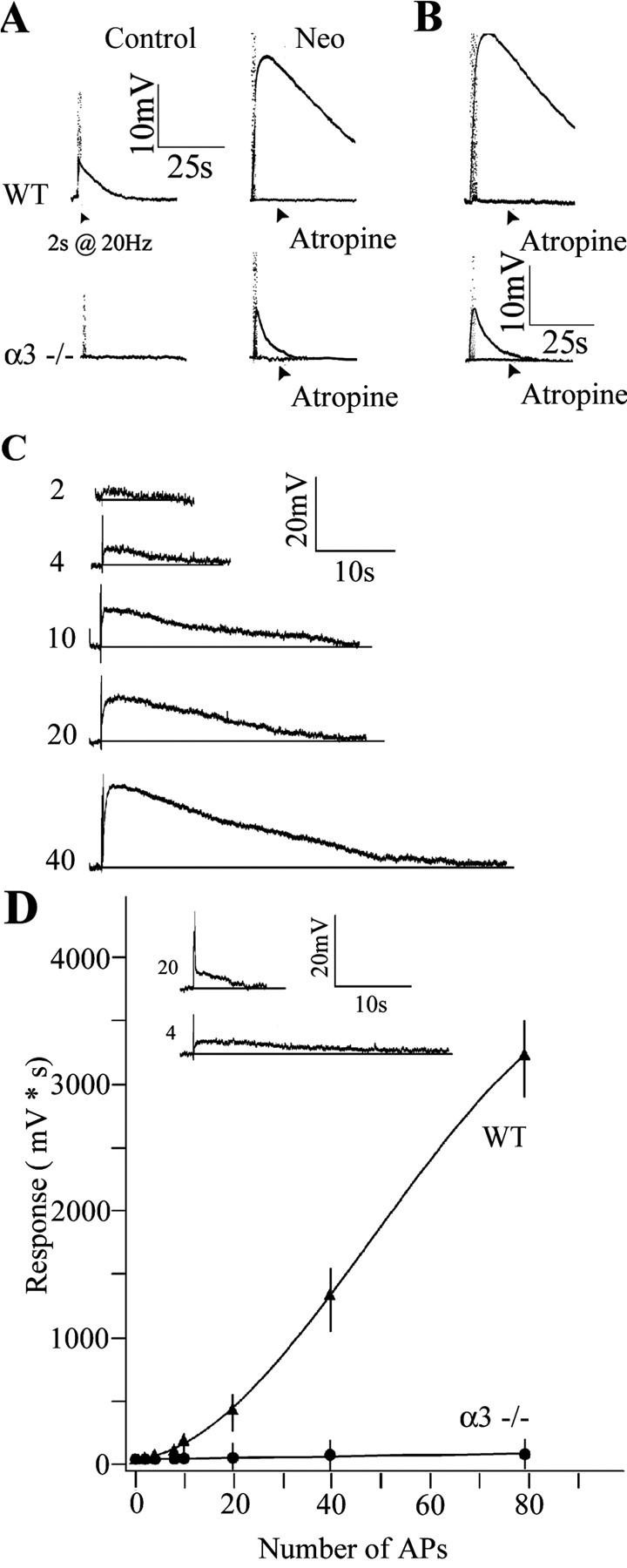
Reduced ACh output from preganglionic varicosities in α3–/– SCG. A, Top traces, Stimulating the preganglionic nerve at 20 Hz for 2 s produced a small, slow muscarinic depolarization on a P6 WT SCG neuron in control solution with hexamethonium (100 μm) (left); the response increased ∼4.5-fold after adding neostigmine (10 μm) (Neo) to inhibit acetylcholinesterase (right) and was blocked by adding atropine (3 μm). Bottom traces, Stimulating the preganglionic nerve at 20 Hz for 2 s produced no detectable response from a P8 α3–/– SCG neuron (left); however, after inhibiting cholinesterase with neostigmine (10 μm) (right), preganglionic nerve stimulation produced a small muscarinic response that was blocked by atropine (3 μm). B, Response from a P21 WT (top) and α3–/– (bottom) SCG neuron after inhibiting cholinesterase with neostigmine (10 μm). C, Muscarinic responses on a P8WT neuron increased with the number of stimuli applied to the preganglionic nerve. The preganglionic nerve was stimulated at 20 Hz; the number of stimuli delivered to the preganglionic nerve is indicated at the left of each trace. D, Integrated muscarinic responses from neurons in WT (▴) and α3–/– (•) ganglia were plotted against the number of applied stimuli to the preganglionic nerve. The solid line represents a fourth-order polynomial fit to the data. The inset compares the response of a P7 WT neuron after four stimuli to the response of a P7α3–/– neuron to 20 stimuli. The values are the means ± SEM (n = 8 for WT and n = 7 for α3–/– neurons). All experiments were done with neostigmine (10 μm) and hexamethonium (100 μm) added to the perfusion fluid at 37°C. AP, Action potential.