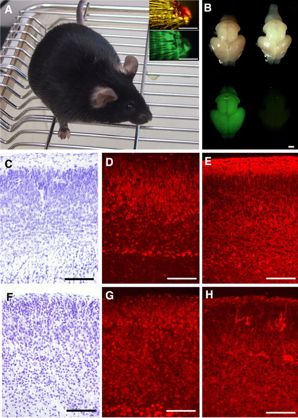
Figure 4.

A mouse cloned from a Cam-2 ES cell, at 6 weeks of age. A, Insets show images of the tail tips photographed using bright-field (top) and fluorescence (bottom) microscopy. B, Gross morphology (top) and GFP-fluorescence (bottom) of the brains of pups on the day of birth. The brains of a pup cloned from Cam-2 ES cells by tetraploid complementation (left) and that of an age-matched normal C57BL/6 mouse (right) are shown. C–H, Sections of the cerebral cortex of a mouse clone from Cam-2ES cells (C–E) and Gad 67-2ES cells (F–H). Nisslstaining (C, F) and indirect immunofluorescence using an antibody to NeuN (D, G) and anti-GAD67 (E, H). No obvious cytoarchitectural deformities were detected. Scale bars: A, B, 1 mm; C–H, 50 μm.
