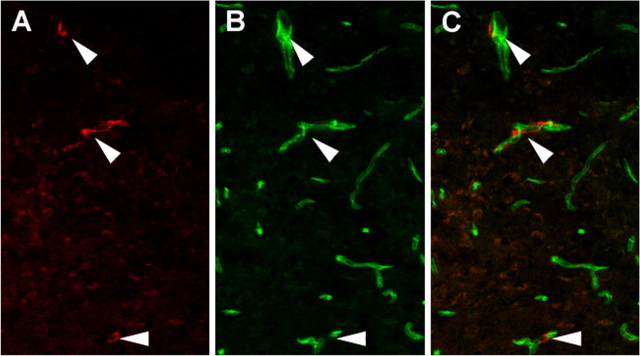
Figure 2.
β-Glucuronidase-positive cells are in close association with the brain vasculature. A-C, Immunohistochemistry for β-glucuronidase (A; red) and PCAM CD31 (B; green), an endothelial cell marker, reveals close association but not overlap (C; merge). Arrowheads denote β-glucuronidase-positive cells.

