Figure 7.
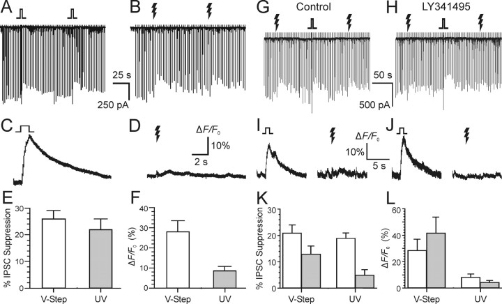
Photoreleased glutamate activates mGluR and induces eCB-dependent responses in cultured hippocampal cells. Each downward deflection represents an evoked GABAA synaptic response. Caged glutamate, Ncm-Glu (500 μm), was in the bath. A, Transient suppression of eIPSCs evoked by 1 s voltage steps to 0 mV on two DSI trials. B, Two trials from the same cell in which 100 ms UV laser flashes were given. C, D, Simultaneous Ca2+ indicator fluorescence measurements for single DSI or glutamate photorelease trials. E, F, Grouped data showing IPSC suppression and Ca2+ transient magnitude from cells treated as in A and B. All data in A-F were obtained from the same six cells. G, Both DSI and glutamate photorelease reduce evoked IPSCs. H, Addition of the mGluR antagonist LY341495 (100 μm) to the bath prevented the photolytic glutamate responses without significantly affecting DSI. I, J, Corresponding Ca2+ signals produced by voltage steps and photoreleased glutamate before (I) and after (J) perfusion of LY341495. K, L, Grouped data (n = 4) for control (open bars) and LY341495 (gray bars) conditions; different cells than A-F. Error bars indicate SE. Jagged arrows represent laser flashes.

