
Figure 1.
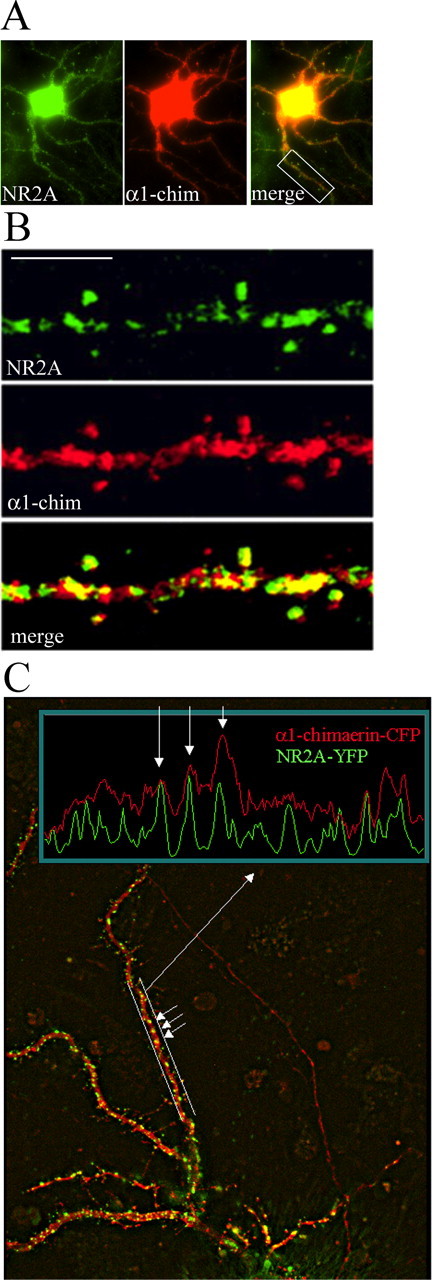
α-Chimerin and NR2A colocalize in hippocampal neurons. A, B, Cultured hippocampal neurons (24 DIV) were cotransfected with α1-chimerin (α1-chim)-CFP fusion and untagged NR2A and stained by immunofluorescence 6 h after transfection for NR2A (green) and CFP (red). The merged image shows colocalization of α1-chimerin and NR2A in dendritic spines. Scale bar, 5 μm. C, Cultured hippocampal neurons were cotransfected with α1-chimerin-CFP fusion and NR2A-YFP and imaged 24 h later. A line profile through a dendritic segment shows that increased α1-chimerin-CFP (red) fluorescence intensity correlates with increased NR2A-YFP (green) fluorescence intensity.