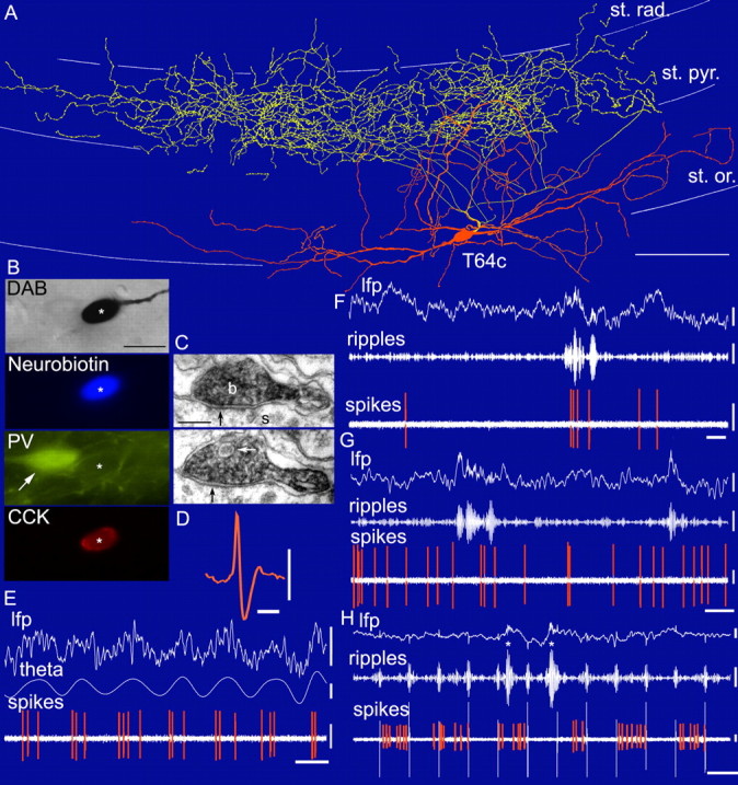
Figure 1.

In vivo firing patterns and visualization of a CCK-expressing basket cell (T64c). A, Reconstruction of the soma and dendrites (orange) is shown complete; the axon (yellow) is shown only from three sections of 65 μm thickness for clarity. st. rad., Stratum radiatum; st. pyr., stratum pyramidale; st. or., stratum oriens. Scale bar, 100 μm. B, Light (DAB reaction) and immunofluorescence microscopic visualization of the labeled cell (asterisk) demonstrates expression of CCK but not PV. Note an adjacent PV-expressing cell (arrow). Scale bar, 20 μm. C, Serial sections of a filled axonal bouton (b) of the cell making a synaptic junction (black arrows) with a pyramidal soma (s). Note the presence of a large granulated vesicle (white arrow), likely to contain neuropeptides. Scale bar, 20 nm. D, Action potential of the recorded cell filtered between 0.8 and 5 kHz. Calibration: 1 ms, 0.5 mV. E-H, In vivo firing patterns of the labeled cell. During theta oscillations (E), the cell fired preferentially at the ascending phase of the theta waves (filtered between 3 and 6 Hz) recorded extracellularly in the pyramidal cell layer by a second electrode. Note the difference in interspike interval between first and second versus second and third spike within a theta cycle (E). During ripple episodes (filtered between 90 and 140 Hz), the cell was sometimes activated (F) and sometimes silenced (G). During the labeling, positive current was applied in 200 ms on/off mode (H), modulating the firing of the cell. Note that a ripple episode during the current-on phase silenced the cell. Asterisks mark true ripple episodes, whereas other fast oscillations are artifacts of current steps. lfp, 0.3-200 Hz. Calibration: lfp, 0.5 mV; theta, 0.2 mV and 0.2 s; ripples, 0.1 mV and 0.2 s; spikes, 0.5 mV.
