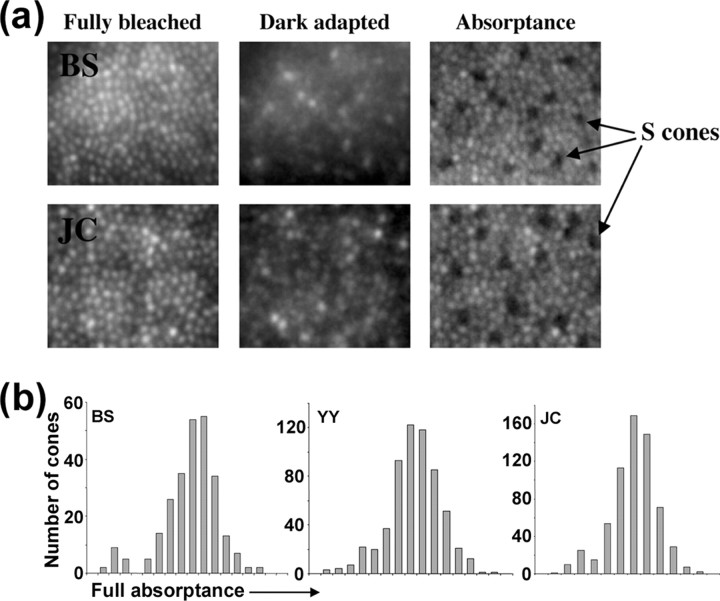
Figure 1.
Identification of S cones in the cone mosaic. a, Averaged retinal images after a full bleach of L- and M-cone pigment, after dark adaptation, and calculated absorptance images. S cones are identified as a sparse array of dim cones in the absorptance image. b, Histogram of individual cone full absorptance values. S cones are identifiable as a low-absorptance peak.

