
Figure 1.
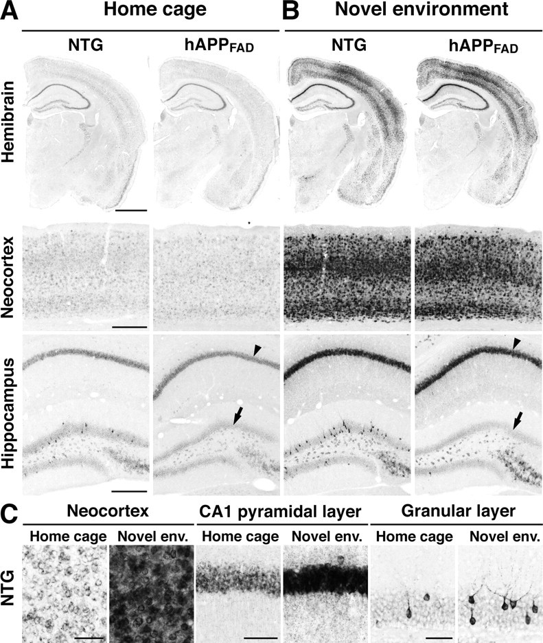
Brain region-specific deficits in basal and stimulated Arc expression in hAPPFAD mice. NTG and TG (hAPPFAD) mice were placed into a novel environment or left in their home cage. Two hours later, they were anesthetized and perfused transcardially. Arc in situ hybridization was performed on floating coronal sections. A, Basal Arc mRNA levels in the hemibrain, neocortex, and hippocampus of mice maintained in the home cage. B, Arc mRNA levels in mice that had explored the new environment. Note the increased Arc expression in multiple brain regions, including the CA1 pyramidal layer (arrowhead), after exploration of the novel environment, and the selective decrease in Arc expression in the granule cell layer of hAPPFAD mice (arrows) under basal (A) and stimulated (B) conditions. C, Higher-magnification images of different brain regions in NTG mice. Novel env., Novel environment. Scale bars: A, B, 1 mm (top), 250 μm (middle, bottom); C, 50 μm.