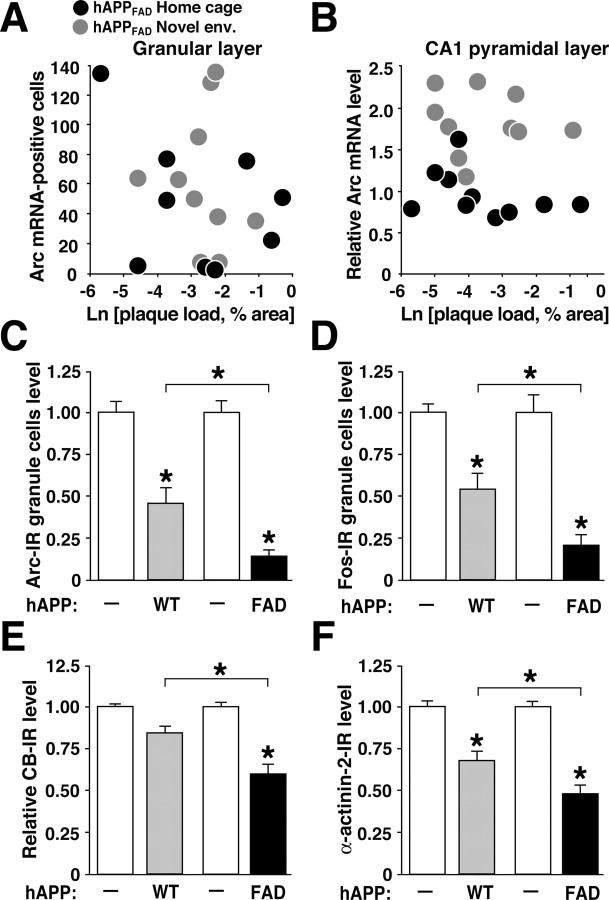
Figure 6.
Arc expression in hAPP mice is independent of early plaque formation but influenced by the extent of Aβ production. A, B, Arc expression in hAPPFAD mice does not correlate with early plaque formation. Coronal brain sections of hAPPFAD mice that had (gray dots) or had not (black dots) explored a novel environment were immunostained for Aβ deposits or analyzed by in situ hybridization to determine the number of Arc-positive granule cells in the dentate gyrus (A) and relative Arc mRNA levels in the pyramidal cell layer of the CA1 region (B). At baseline and after exploration, neuronal Arc expression in the granular layer and in CA1 did not correlate with the extent of amyloid deposition in these regions. C-F, Molecular profile of granule cells in two hAPP lines with similar levels of hAPP expression but different levels of Aβ (Mucke et al., 2000). Brain sections from TG mice of hAPPWT line I5 ( ) and hAPPFAD line J20 (▪) and from NTG littermates of each line (□) were immunostained for Arc (C), Fos (D), calbindin (E), or α-actinin-2 (F). n = 9-12 mice per group; *p < 0.01 (Tukey-Kramer test). Error bars indicate SEM. Ln, Natural log.

