
Figure 1.
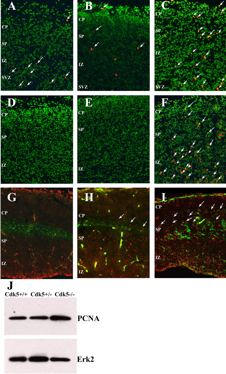
Loss of Cdk5 leads to loss of cell cycle control in E16.5 neocortex. A-C, Coronal sections of Nissl-stained neocortex (green) shows the location and number of BrdU-positive neurons (red) in Cdk5+/+ (A), Cdk5+/- (B), and Cdk5-/- (C)embryos. Arrows denote BrdU-positive cells. D-F, Coronal sections labeled with anti-PCNA (red) in Cdk5+/+ (D), Cdk5+/- (E), and Cdk5-/- (F) cortex. Labeled cells are apparent in the subventricular zone (SVZ), intermediate zone (IZ), subplate (SP), and cortical plate (CP) of the Cdk5-/- brain. Arrows denote examples of PCNA-positive cells. G-I, Sagittal sections labeled with anti-chondritin sulfate proteoglycan (green) and anti-Ki67, a proliferation marker (red). The Cdk5-/- cortex (I) has many Ki67-positive cells in both SP and CP. In the wild-type cortex (G), there are no positive cells in or above the subplate, and, in the heterozygote cortex, there is an intermediate phenotype with some Ki67-positive cells in the subplate (H). J, Western blot of an E16.5 neocortical lysate shows an increase in the PCNA protein content in the Cdk5-/- brain (lane three).