Figure 4.
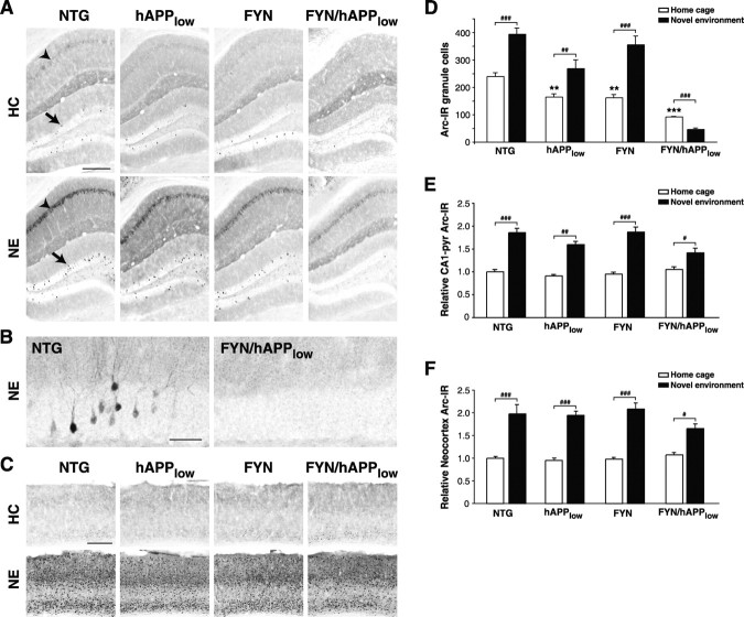
Impaired induction of Arc expression in FYN/hAPPlow mice. A, Micrographs demonstrating Arc-IR granule cells in the dentate gyrus (arrow) and pyramidal cells in CA1 (arrowhead) in home-caged mice (HC; top) and mice that had explored a novel environment (NE; bottom). B, High-magnification micrographs illustrating that granule cells in the dentate gyrus were Arc immunoreactive in an NTG mouse (left), but not in a FYN/hAPPlow mouse (right), after exploration of a novel environment. C, Micrographs demonstrating Arc-IR cells in the neocortex of mice that had (bottom) or had not (top) explored the novel environment. D-F, Quantitation of Arc immunolabeling (n = 3-10 mice per genotype and condition) demonstrates that environmental exploration increased the number of Arc-positive granule cells in NTG and singly TG mice but not in FYN/hAPPlow mice (D). In contrast, environmental exploration increased Arc induction in the pyramidal (Pyr) cell layer of the CA1 region (E) and the neocortex (F) also in FYN/hAPPlow mice, although to a somewhat lesser extent than in NTG and FYN mice. For CA1 and neocortex data, measurements in each bar were normalized against the mean of the home-caged NTG mice. **p < 0.01, ***p < 0.001 versus NTG by ANOVA; ##p < 0.01, ###p < 0.001 by Fisher's PLSD post hoc test. Scale bars: A, C, 250 μm; B, 50 μm.

