
Figure 2.
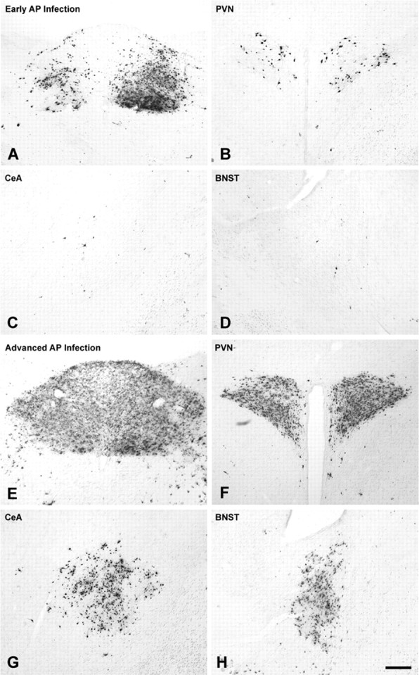
Immunocytochemical localization of PRV-positive neurons in control NH/NS rat pups injected with virus on P8 and killed after 63-66 h, on P10. The magnitude of forebrain infection (PVN, BNST, CeA) in individual cases correlated with the extent of infection in the AP (Table 2). Forebrain infection was sparse in cases that exhibited robust infection of the left DMV and NST but only scattered infection of AP (A-D). In contrast, extensive infection of neurons in the PVN, BNST, and CeA invariably accompanied extensive AP infection (E-H). Scale bar: (in H) A-H, 100 μm. See Figure 1 for regional schematics.