
Figure 4.
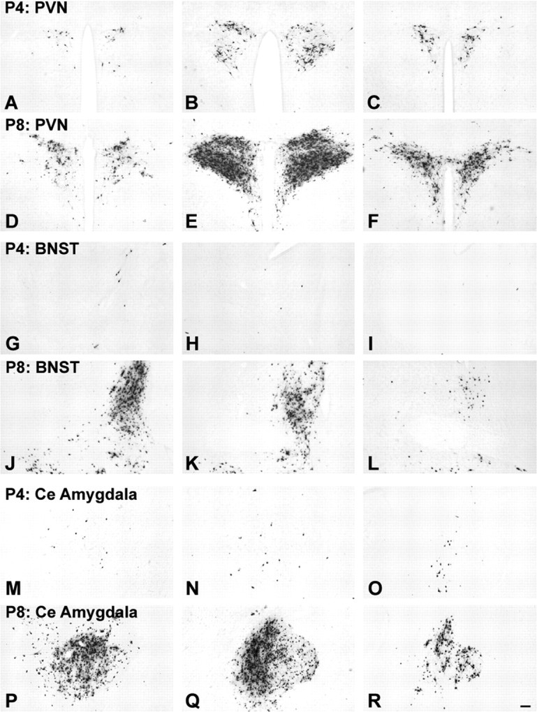
Age-dependent assembly of preautonomic circuits. Large numbers of transneuronally infected neurons are evident in the PVN (D-F), BNST (J-L), and CeA (P-R) in control NH/NS rat pups injected with virus on P8 and killed on P10; evidence that these synaptic inputs from neurons in these regions to gastric autonomic output neurons are well established by this time. In contrast, significantly fewer infected neurons are evident within the PVN (A-C), BNST (G-I), or CeA (M-O) in NH/NS rat pups injected with virus on P4 and killed on P6, despite comparable infection within the hindbrain DVC in all cases (see also Fig. 3). Each row of photomicrographs depicts sections through rostral (left), intermediate (middle), and caudal (right) levels through each region. Scale bar: (in R) A-R, 100 μm. See Figure 1 for regional schematics.