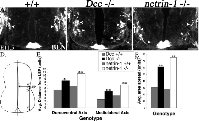
Figure 5.
SACMN cell bodies fail to migrate dorsally, and some of their axons inappropriately extend toward the floor plate, in both Dcc and netrin-1 null embryos. A-C, Anti-BEN labeling of transverse, cervical spinal cord-containing cryosections derived from E11.5 wild-type and Dcc or netrin-1 null embryos. Anti-BEN selectively labels small clusters of SACMNs (arrowheads), which are appropriately positioned just beneath the LEP, as well as the SAN (arrows) in an E11.5 wild-type embryo (A). In a Dcc null littermate (B), anti-BEN labels disorganized clusters of SACMN cell bodies, many of which have failed to migrate to, and settle within, the vicinity of the LEP (arrowheads). Similarly disorganized anti-BEN-positive SACMN cell bodies, as well as a SACMN axon that appears to be inappropriately projecting toward the floor (arrowheads), are present in a netrin-1 null E11.5 embryo (C). D-F, Analysis and quantification of the defects observed in Dcc and netrin-1 mutant embryos. D, A schematic representation of the spinal cord indicating that SACMN/axon migration defects were scored along the D-V axis between the LEP and the ventral margin of the spinal cord, as well as along the M-L axis, between the LEP and the central canal. E, BEN-positive SACMNs were detected at significantly greater distances from the LEP in Dcc and netrin-1 mutants compared with their wild-type littermates, along both the dorsoventral and mediolateral axes. F, The area spread (product of average dorsoventral and mediolateral distances) of BEN-positive SACMNs was significantly greater in Dcc and netrin-1 mutants than in their wild-type littermates. Values shown are means; *p < 0.05, **p < 0.005. 1 unit is equivalent to 20 μm. B, n = 11; C, n = 4. Scale bar, 100 μm.

