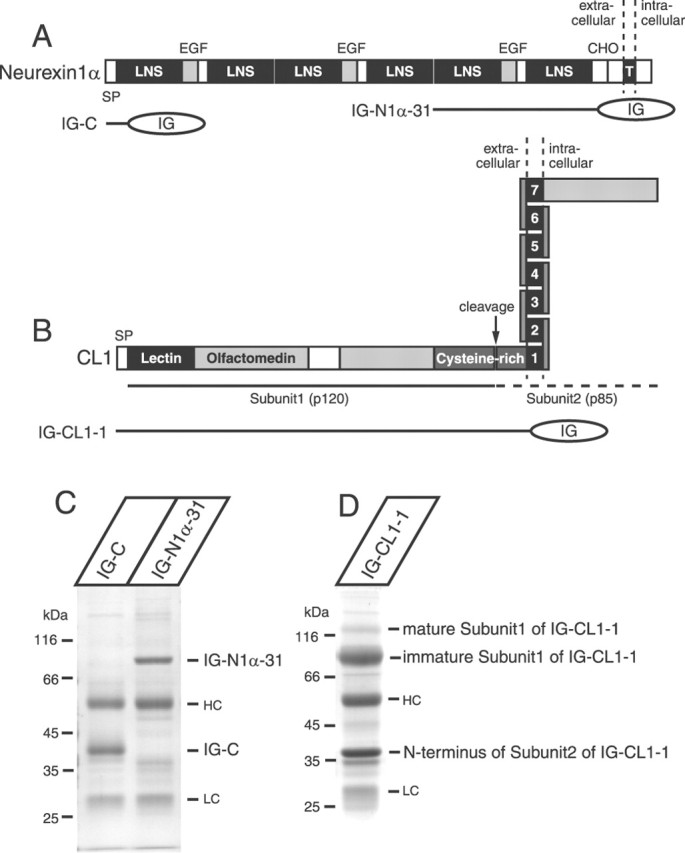
Figure 3.

Analysis of purified recombinant neurexin 1 α and CL 1 proteins expressed in COS-7 cells. A, Domain structure of neurexin 1α and location of Ig fusion proteins used for binding to α-latrotoxin. Principal features of neurexin 1α are indicated on the top as follows: SP, signal peptide; LNS, LNS domains; EGF, epidermal growth factor-like sequences; CHO, carbohydrate attachment site; T, transmembrane region. B, Domain structure of CL1 and location of Ig fusion protein used for binding to α-latrotoxin. SP, signal peptide; lectin, lectin-like domain; olfactomedin, a domain homologous to olfactomedins and myocilin. Close to the membrane is a cysteine-rich domain that contains a cleaving site. C, Purified recombinant neurexin 1α (the fifth and the sixth LNS domains; IG-N1α-31) was analyzed with the control protein (IG-C) by SDS-PAGE and Coomassie staining. D, Analysis of the recombinant, entire extracellular domain of CL1 (IG-CL1-1) by SDS-PAGE and Coomassie staining. HC, Heavy chain; LC, light chain.
