Figure 2.
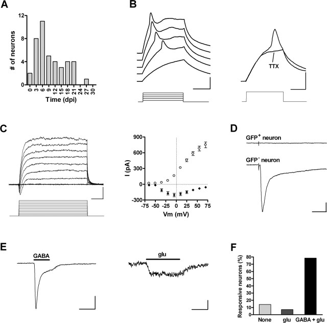
Electrophysiology of silent neurons. A, Age of GFP+ neurons exhibiting action potentials and lacking synaptic inputs (n = 42). B, Left, Depolarizing current steps (10 pA) elicit short and wide action potentials in a 5 dpi neuron. Right, Immature spikes are blocked by 1 μm TTX (step, 20 pA). Calibration: 20 mV, 20 ms. C, Left, Depolarizing voltage steps (15 mV) elicit fast inward and slow outward currents in a 4 dpi neuron. Calibration: 200 pA, 5 ms. Right, I-V plot of peak inward current (filled diamonds) and steady-state outward current (open circles). Data are mean ± SEM (n = 35). D, Lack of postsynaptic responsiveness of a GFP+ neuron (top trace) to extracellular stimulation of the GCL (30 V, 50 μs). A neighboring GFP- neuron did exhibit a complex inward current in response to the same stimulus (bottom trace). Calibration: 50 pA, 30 ms. E, Responses of a 4 dpi neuron to focal application of 0.5 mm GABA (Ipeak, -98.6 ± 24.5 pA; n = 11; left) and 0.5 mm glu (Ipeak, -12.1 ± 2.5 pA; n = 11; right). Calibration: 50 pA (left), 5 pA (right), 10 s. F, Percentage of GFP+ neurons responding to exogenous application of GABA and glu (n = 14 cells).

