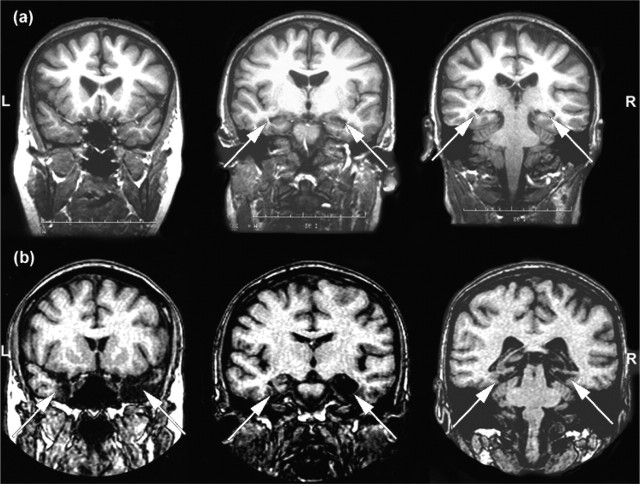
Figure 1.
Three coronal MRI scan slices for one representative patient from the hippocampal (a) and MTL (b) patient groups are shown (arrows highlight regions of significant damage). L, Left; R, right. Reproduced with permission from Lee et al. (2005a).

