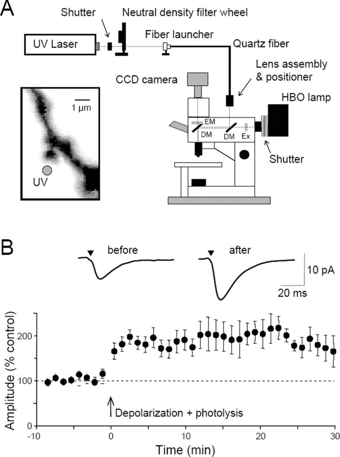
Figure 1.
Potentiation of exogenous glutamate responses at single dendritic spines. A, Diagram of the experimental apparatus. UV light is launched into a quartz multimode fiber and focused in a conjugate focal plane using a lens assembly. The intensity of the light is controlled by a neutral density filter wheel. A dichroic mirror (DM) allows the UV light to be combined with wide-field illumination from a conventional mercury arc (HBO) lamp, after passing through an excitation filter (EX). The normal filter cube contains a second dichroic mirror and the desired emission filter (EM). Mechanical shutters gate the UV and visible light pulses. A CCD camera captures images of the cells. Inset, With this apparatus, UV light for microphotolysis can be directed to a spot (gray circle) near the head of a single dendritic spine, visualized with fluorescent dye loaded into the cell from the whole-cell patch pipette. These spots have a full-width at half-maximal height of <1 μm. B, Pooled data showing the mean ± SEM potentiation of photolytic EPSCs in 13 cells. Averaged photolytic EPSCs 3 min before and 20 min after the pairing are shown above and were elicited from the spine illustrated in A. Modified from Bagal et al. (2005).

