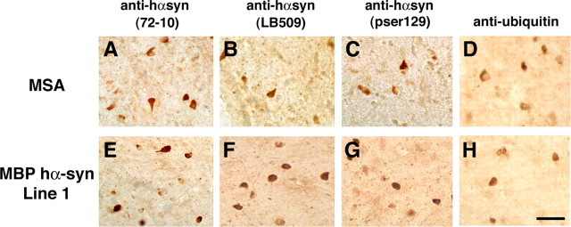
Figure 3.
Comparison of the glial cell inclusions between MSA and MBP hα-syn tg animals. Images are from the white matter tracts in the basal ganglia of a human case with typical MSA and MBP hα-syn tg mice from line 1 (4 months of age). A, Conical and ovoid GCIs in an MSA case were positively immunostained with a polyclonal antibody against hα-syn (72-10). B, GCIs immunostained with a monoclonal antibody against hα-syn (LB509). C, GCIs immunostained with a monoclonal antibody against phospho-serine129 hα-syn (pser129). D, GCIs immunostained with an antibody against ubiquitin. E, Conical and ovoid glial inclusions in an MBP hα-syn tg mouse were positively immunostained with a polyclonal antibody against hα-syn (72-10). F, Glial inclusions immunostained with a monoclonal antibody against hα-syn (LB509). G, Glial inclusions immunostained with a monoclonal antibody against phospho-serine129 hα-syn (pser129). H, Glial inclusions in the tg mice were mildly immunostained with an antibody against ubiquitin. Scale bar, 20 μm.

