Figure 5.
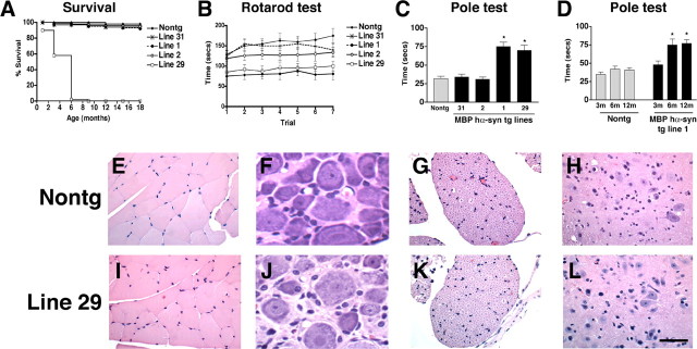
Characterization of survival and neurological deficits in MBP hα-syn tg mice. A, Survival curves for the MBP hα-syn tg mice showing that, although mice from the lower-expresser lines (1, 2, and 31) were viable for long periods of time, mice from high-expresser line 29 died prematurely by 6 months of age. B, Motor assessment in the rotarod showed that compared with nontg mice (n = 6), tg mice from line 31 (n = 6) had a mild impairment of motor function, whereas tg mice from lines 2 (n = 6), 1 (n = 6), and 29 (n = 6) had more significant motor deficits in this test. Mice were tested at 4 months of age. C, Motor assessment in the pole test showed that compared with nontg mice (n = 6), tg mice from lines 31 (n = 6) and 2 (n = 6) demonstrated motor abilities comparable with control mice, whereas tg mice from higher-expresser lines 1 (n = 6) and 29 (n = 6) displayed significant motor impairment. Mice were tested at 4 months of age. D, Motor assessment in the pole test showed that, at 3 months of age, only mild deficits were observed; however, at 6 months of age, these deficits were accentuated and remained similar at 12 months of age. Error bars are mean ± SEM. *Significant difference compared with nontg controls (p < 0.05; one-way ANOVA with post hoc Dunnett's). E-L, For histological analysis, paraffin sections from 4-month-old mice were stained with hematoxylin/eosin and imaged by bright-field microscopy. E-H, Histological analysis of the hindlimb muscle (E), dorsal root ganglion (F), spinal nerve roots (G), and motor neurons (H) in the thoracic segments of the spinal cord in nontg mice. I-L, No significant alterations were observed in muscle (I), dorsal root ganglion (J), spinal nerve roots (K), and motor neurons (L) in the thoracic segments of the spinal cord in MBP hα-syn tg mice. Scale bar, 20 μm.

