
Figure 8.
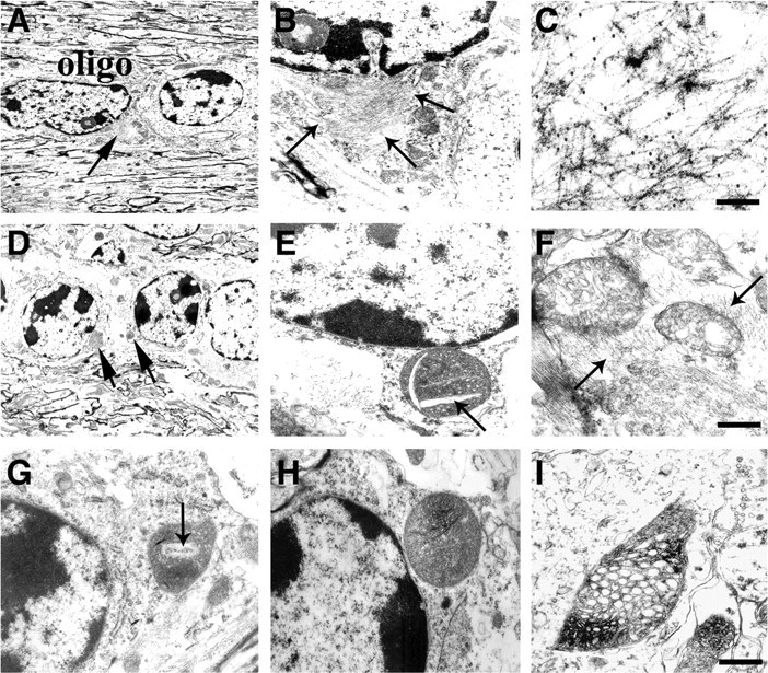
Ultrastructural analysis of the neuropathological alterations in MBP hα-syn tg mice. All panels are from the neocortex of 4-month-old tg mice from line 29 imaged by electron microscopy. A, B, Oligodendroglial cells (oligo) within the white matter tracts in the corpus callosum contain fibrillary perinuclear inclusions (arrows), which are surrounded by electrodense material. C, Immunoelectron microscopic analysis of the oligodendroglial cells with an antibody against hα-syn shows abundant gold particles labeling the filaments in the inclusions. D-F, The mitochondria within oligodendroglial cells display abnormal characteristics, including increased size (D), crystalline-like inclusions (arrows; E), irregular crista, and the accumulation of filaments (arrows) around the mitochondria (F). G-I, Mitochondrial alterations in neuronal cells, including formation of crystalline-like inclusions (arrow; G), increased size (H), and irregular crista with electrodense material (I). Scale bars: (in C) A-E, G, H, 10 μm; F, I, 1 μm.