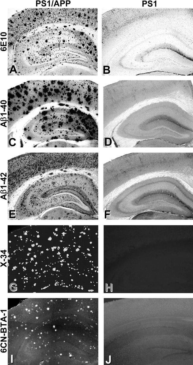
Figure 3.

Histological analysis shows extensive amyloid deposition in the PS1/APP mice but not the PS1 mice. Tissue sections from PS1/APP (A, C, E, G, I) and PS1 (B, D, F, H, J) mice were immunostained with antibodies to total Aβ (A, B; 6E10), Aβ1-40 (C, D), and Aβ1-42 (E, F). Sections also were stained with the fluorescent Congo red derivative X-34 (G, H), which stains fibrillar amyloid, and the fluorescent PIB analog 6-CN-BTA-1 (I, J).
