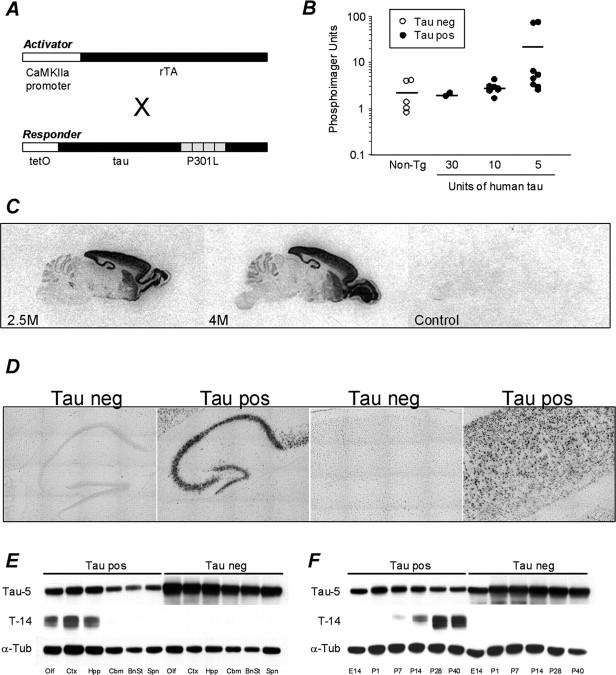
Figure 1.
Generation and characterization of rTg(tauP301L)4510 mice. A, Diagrammatic representation of activator and responder transgenes in bigenic mice. B, Highest levels of tauP301L expression were achieved in mice harboring the lowest number of responder transgene copies. C, Specific transgene expression in the forebrain was confirmed by in situ hybridization in 2- and 4-month-old rTg(tauP301L)4510 mice. Emulsion-dipped slides revealed silver grains clustered over individual neurons, of tau-positive mice only, indicating expression was restricted to neuronal cells types and that the majority of neuronal cells in the hippocampus and cortex expressed transgenic transcripts. D, Composite images (16 images at ×20) of emulsion-dipped slides from 4-month-old tau-positive and tau-negative mice. E, Biochemical studies confirmed high expression of tauP301L in the hippocampus and cortex and no detectable expression in the spinal cord of 2.5-month-old mice. F, Western blot analysis of postnatal forebrain indicated the earliest expression of transgenic tauP301L in P7 mice. Mouse and human tau were detected with tau-5 antibody (Biosource), and human-specific tau was probed for with T14 (Zymed). To demonstrate the high levels of transgene expression, tau-5 immunoblots were loaded with 3 μg of tau-positive forebrain extract compared with 30 μg of tau-negative lysate. T14 and α-tubulin blots were run in parallel after equal loading of 3 μg of protein across all lanes. neg, Negative; pos, positive; Tg, transgenic;α-Tub,α-tubulin; Olf, olfactory bulb; Ctx, cortex; Hpp, hippocampus; Cbm, cerebellum; BnSt, brainstem; Spn, Spinal cord.

