Figure 4.

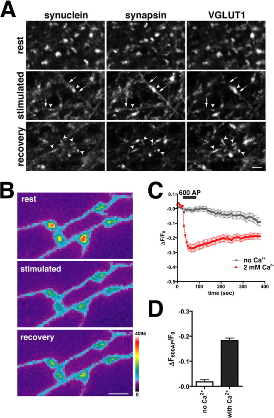
α-Synuclein disperses in response to depolarization. A, Neurons were fixed (rest), depolarized with 45 mm KCl and immediately fixed (stimulated), or depolarized followed by a 10 min recovery (recovery) before fixation. Synaptic boutons were identified by VGLUT1 staining and are indicated with arrowheads. Similar to synapsin, endogenous α-synuclein disperses from boutons after depolarization. Unlike synapsin, however,α-synuclein does not accumulate in the axon (arrows) after stimulation. Ten minutes after recovery, synapsin has reclustered in the synaptic terminal and colocalizes with VGLUT1. α-Synuclein does not reaccumulate at the synapse in this time frame. Scalebar, 2 μm. B, At rest, GFP-α-synuclein is enriched at synaptic boutons and disperses during stimulation with 600 action potentials (AP) delivered at 10Hz. Minimal reclustering of the protein has occurred by 5 min (recovery). The color scale is shown to the right. Scalebar, 2 μm. C, No dispersion of α-synuclein occurs during stimulation with 600 AP in calcium-free medium (gray). After stimulation in the absence of calcium, the cells were washed in calcium-containing medium for 10 min and were then restimulated (red). The dispersion of α-synuclein thus depends on calcium entry. The traces are the average ± SEM dispersion at 30 synapses from one representative cell. D, Average dispersion of GFP-α-synuclein during sequential stimulation in calcium-free and calcium-containing medium. Medium was exchanged during a 10 min rest separating the two stimulation rounds. Error bars indicate SEM. p<0.0001, Student's ttest; n = 138 synapses from three cells.
