
Figure 5.
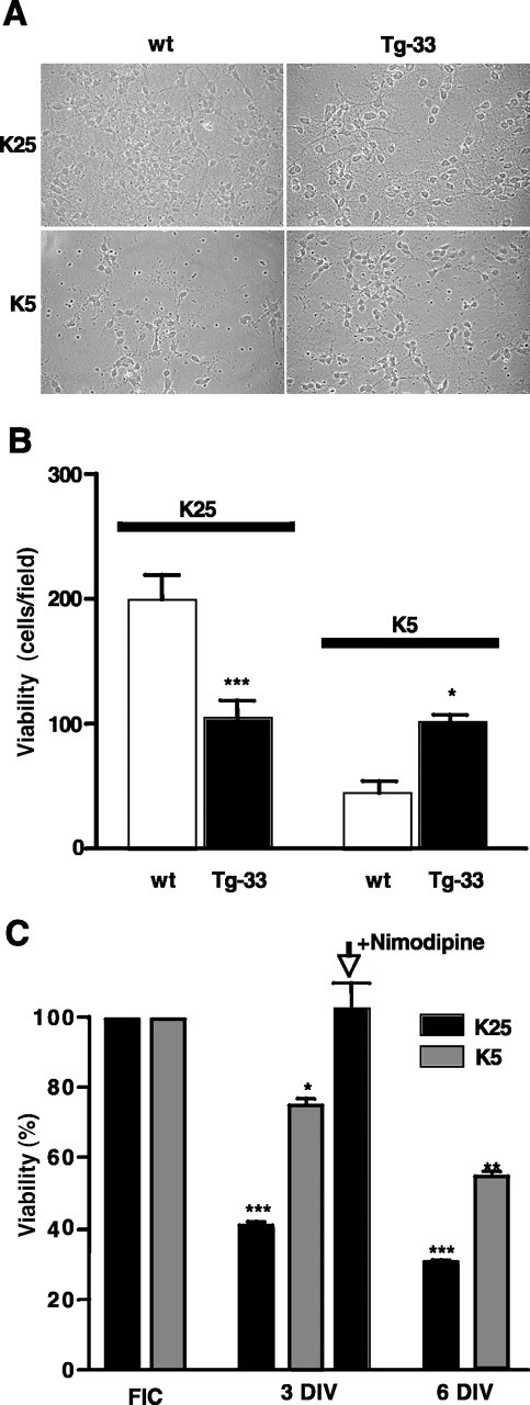
Differential effects of extracellular potassium concentrations on the viability in EFmDREAM transgenic neurons. A, Phase-contrast light micrographs of cerebellar neurons from wild-type (wt) or Tg-33 mice cultured for 3 d in vitro (3 DIV) in media containing either 25 or 5 mm KCl. B, Quantitation of cerebellar granules from wild-type or Tg-33 transgenic mice after 3 d in culture using 4′,6′-diamidino-2-phenylindole dihydrochloride (DAPI) staining. The bars represent the mean ± SEM of the total number of DAPI-labeled cells in different fields. Six different fields in four different cultures were counted. *p < 0.05; ***p < 0.001, one-way ANOVA test. C, Quantitation of the viability of transgenic granules estimated indirectly by real-time reverse transcription-PCR of EFmDREAM mRNA in cultured Tg-33 cerebellar granules maintained under depolarizing (25 mm KCl) or basal (5 mm KCl) conditions. The results from freshly isolated cells (FIC) and from cells at 3 and 6 DIV correspond to the mean ± SEM of six experiments using different cultures. Significant differences from control (EFmDREAM expression in FIC) are indicated as *p < 0.05, **p < 0.01, and ***p < 0.001 (one-way ANOVA).