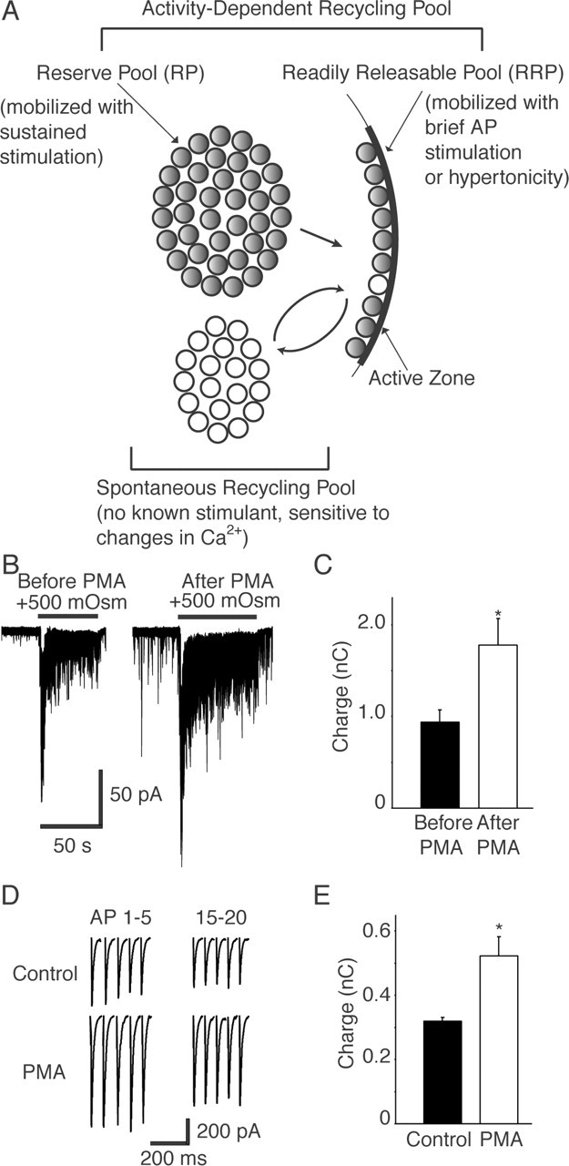
Figure 2.

A, Studies in several secretory preparations and central neuronal synapses have classified synaptic vesicles with respect to their relative availability for release during stimulation. The vesicles in the RRP are immediately available for release after arrival of an action potential. RRP vesicles can be mobilized by a brief burst of action potentials or by application of hypertonic solutions (Rosenmund and Stevens, 1996). A reserve pool (RP) of vesicles is spatially distant from the release sites and replaces the vesicles in the RRP that have undergone exocytosis. RP vesicles can typically be mobilized by sustained trains of action potentials or by elevated K+ stimulation. Together, the RRP and reserve pool make up the recycling pool of vesicles, which corresponds to all vesicles capable of activity-dependent recycling (all gray vesicles) after stimulation. Recent evidence suggests that spontaneous neurotransmission is driven by a set of vesicles recycling independently of the activity-dependent vesicle pool (white vesicles) (Sara et al., 2005). Note that a comparison of the total number of vesicles measurable in synapses through physiological means versus morphological analysis supports the presence of a number of vesicles that are not mobilized by activity under standard paradigms. The mechanisms that would fully mobilize this “resting” pool of vesicles (not depicted in the figure) remain to be determined. B, PMA application increased the size of the RRP in 15 DIV hippocampal cultures. Sample whole-cell patch-clamp recordings of synaptic responses to the application of 500 mm sucrose solution before and 2 min after PMA treatment are shown. C, Plot showing an increase in the size of sucrose response after PMA treatment, measured as the total charge (n = 3). *p < 0.05. D, Sample traces of whole-cell patch-clamp recordings showing the first five and last five responses to a 20 Hz, 2 s train of action potentials recorded before and after application of PMA in 15 DIV neuronal cell cultures. E, Plot showing the PMA-dependent increase in total synaptic charge transfer in response to 20 Hz, 2 s electrical stimulation compared with controls (n = 5). *p < 0.05. Error bars indicate SE.
