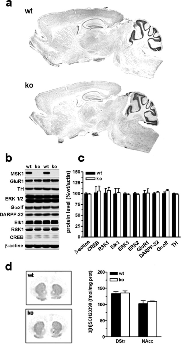
Figure 3.

Brain morphology and protein expression in MSK1 knock-out mice. a, Brain morphology was analyzed by using cresyl violet staining in wild-type (wt) and MSK1-KO (ko) mice. b, Expression levels of some important proteins for cell signaling studied by immunoblotting in striatal extracts. MSK1 is expressed strongly in the striatum of wild-type mice. None of the analyzed proteins was modified in MSK1-KO mice. c, Quantification of protein expression levels in wt and ko mice (means ± SEM; n = 7 mice per group). d, D1 receptor binding was studied and quantified by using [3H]SCH23390 as a radioligand (means ± SEM; n = 3 mice per group).
