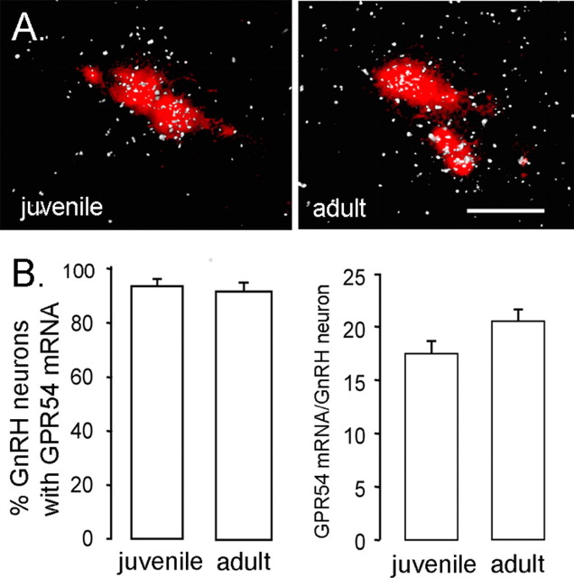
Figure 4.
Expression of GPR54 mRNA in GnRH neurons across development. A, Representative photomicrographs of cells coexpressing GPR54 mRNA (reflected by silver grains, appearing as clusters of white dots) and GnRH mRNA (labeled with Vector Red) in juvenile (left) and intact adult (right) male mice. Scale bar, 20 μm. B, Quantitative analysis of GPR54 mRNA in GnRH neurons demonstrated that neither the percentage of double-labeled cells (left) nor the relative expression of GPR54 mRNA in GnRH neurons (reflected by the number of silver grains per GnRH neuron; right) differed significantly between P18 and adult mice (n = 6/7 per group). Errorbars represent SEM.

