Figure 5.

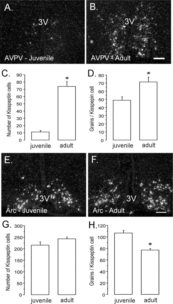
Developmental changes in KiSS-1 mRNA expression within the anteroventral periventricular and arcuate nuclei. A–D, KiSS-1 mRNA in the AVPV. Photomicrographs show AVPV cells possessing KiSS-1 mRNA (reflected by silver grains, appearing as clusters of white dots) in juvenile (left) and adult (right) male mice. Quantitative analysis of kisspeptin mRNA revealed that both cell number (C; p < 0.0001) and cell content (D; p < 0.05) of kisspeptin mRNA increased significantly from juvenile to adult mice (n = 6/7 per group). E, F, KiSS-1 mRNA in the arcuate nucleus (Arc). Photomicrographs show KiSS-1 mRNA in the arcuate nucleus (Arc) of juvenile (E) and adult (F) male mice (n = 6/7 per group). Quantitative analysis of KiSS-1 mRNA in the Arc revealed that the number of positive cells did not differ between juvenile and adult mice (G); however, the per cell content of KiSS-1 mRNA (reflected by the number of silver grains per cell) was reduced in adult mice (H; p < 0.001). The asterisks denote statistical significance. Scale bars, 100 μm. 3V, Third ventricle. Error bars represent SEM.
