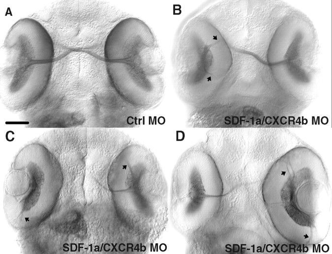
Figure 4.
Retinal axons make pathfinding errors as a result of reduced SDF-1a/CXCR4b signaling after MO knockdown. All panels are ventral views of 48 hpf embryos labeled with MAb Zn5, which stains both RGC cell bodies and axons. A, The optic nerve and RGCs labeled by MAb Zn5 in an embryo injected with control MO. B-D, RGC axons frequently projected aberrantly (arrows) in embryos injected with sdf-1a and cxcr4b antisense MOs. In some cases (left-side retinas in B and C), no optic nerve formed with all MAb Zn5+ retinal axons projecting abnormally within the eye. Scale bar, 20 μm.

