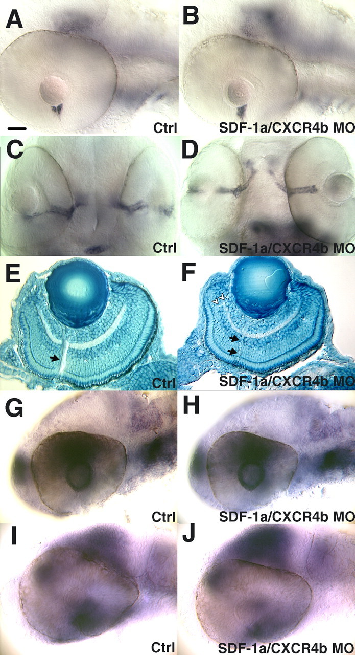
Figure 5.

Perturbation of SDF-1/CXCR4 expression does not affect the patterning of the retina and optic stalk, nor does it change the expression pattern of other guidance molecules. A, Expression of netrin-1a by the head of the optic stalk adjacent to the eye is normal after heat induction as seen in a side view of a 48 hpf control MO-injected embryo. Anterior is left; dorsal is up. B, Expression of netrin-1a is unperturbed after antisense SDF-1a and antisense CXCR4b MO injection. C, Expression of pax2a by the optic stalk seen in a ventral view of a 48 hpf embryo injected with control MO. D, Expression of pax2a is unperturbed after knockdown of SDF-1a/CXCR4b. E, Sagittal section of the eye stained with methylene blue, showing the architecture of a 72 hpf embryo after injection of control MOs. The arrow denotes the optic axons exiting the retina. F, Knockdown of SDF-1a and CXCR4b does not disturb the architecture of the eye. Note the thin bundle of optic axons exiting the retina (arrow) and the aberrant axon tract (arrowheads). G, Side view of a control MO injected 32 hpf embryo showing the expression of ephrinB2a by cells in the lens and the dorsal retina. H, Knockdown of SDF-1a and CXCR4b does not alter eprhinB2a expression. I, Side views of control or MO-injected embryos at 32 hpf showing ephB3 expression in the nasal region of the retina as well as the tectum. The apparent expression seen in the dorsal retina in I and in J is actually expression by the brain just medial to the eye. J, ephB3 expression was not disturbed by SDF-1a/CXCR4b knockdown. Scale bar, 20 μm.
