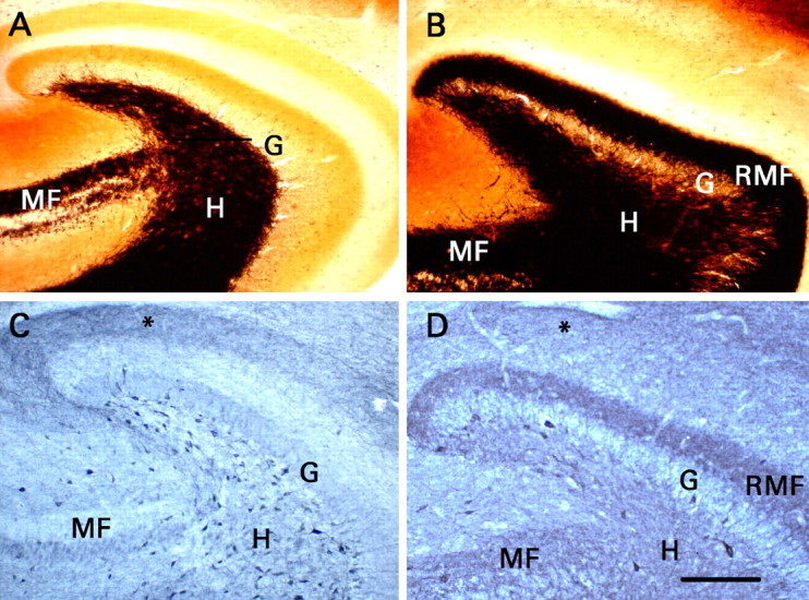
Figure 1.

Recurrent mossy fibers that expressed NPY were present when electrophysiological studies were performed. Transverse sections were cut from the same portion of the hippocampal formation used for recording. G, Granule cell body layer; H, dentate hilus; MF, mossy fiber pathway in area CA3b-CA3c; RMF, recurrent mossy fiber pathway. A, Timm histochemistry in a section from a control rat. Black mossy fiber-like staining is present only in the mossy fiber pathway of area CA3 and the dentate hilus. B, Timm histochemistry in a section from a rat that had developed status epilepticus and did not receive phenobarbital. Mossy fiber-like Timm staining is present in the inner third of the dentate molecular layer, where recurrent mossy fibers innervate granule cell dendrites. C, NPY immunohistochemistry in a section from a control rat. Note the presence of numerous immunoreactive somata in the dentate hilus, terminal immunoreactivity in the outer part of the dentate molecular layer (asterisk), and little immunoreactivity either in the mossy fibers of area CA3 or in the inner third of the dentate molecular layer. D, NPY immunohistochemistry in a section from a rat that had developed status epilepticus and did not receive phenobarbital. Note the neoexpression of NPY in the mossy fibers of area CA3b-CA3c, in the hilus, and particularly in the recurrent mossy fibers. Status epilepticus reduced the number of hilar NPY-immunoreactive somata markedly, and terminal immunoreactivity in the outer part of the molecular layer (asterisk) was less distinct than in C. C, D, Staining of granule and pyramidal cell bodies resulted from nonspecific binding of the secondary antibody and does not signify expression of NPY. Scale bar (in D) A-D, 200 μm.
