Figure 2.
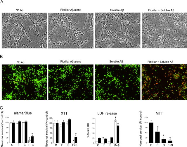
The presence of both soluble Aβ and fibrillar Aβ are required for Aβ-mediated neuronal cell death. Cultures of human cortical neurons were treated with media alone or media containing 1 μm fibrillar Aβ. After 1 h, culture medium was aspirated and replaced with media alone or media containing 20 μm soluble Aβ. In total, four treatment conditions were used in this study: pretreatment with media alone followed by media-alone treatment, which was used as the control condition (“C”); pretreatment with fibrillar Aβ, followed by media-alone treatment (“F”); pretreatment with media alone, followed by soluble Aβ treatment (“S”); and pretreatment with fibrillar Aβ, followed by soluble Aβ treatment (“F + S”). Primary neuronal cultures were incubated for 3 d and then assayed for neuronal viability by phase contrast microscopy (A), live cell (green) and dead cell (red) staining (B), and by measuring cellular redox activity using alamarBlue, the tetrazolium redox dye XTT that forms a soluble formazan product, and the tetrazolium redox dye MTT that forms an insoluble formazan product (C). C, Third panel, The integrity of the neuronal cell membrane was quantitatively determined by measuring the release of the cytoplasmic enzyme LDH into the culture medium. Total LDH in cells was determined by lysing cells with 1% Triton X-100. This value was used to calculate percentage of total LDH released. The white and black bars represent data obtained using independent culture preparations. The modest effect of fibrillar Aβ on LDH release obtained in one experiment (white bars), although significant, was not repeated in additional studies using fibrillar Aβ. Error bars indicate ± SD. *p < 0.001 versus control cultures. Statistical analysis was based on the means ± SD (n = 6) using ANOVA, followed by Fisher's PLSD test using StatView software (version 5.0; SAS Institute, Cary, NC).

