
Figure 4.
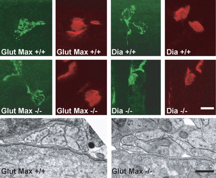
The development of NMJs is not changed in gephyrin-deficient mice at P0. The top two rows show confocal images of NMJs from wild-type (+/+) and gephyrin-deficient (-/-) mice that have been stained for axon nerve terminals with anti-neurofilament plus anti-synaptophysin (green) and for acetylcholine receptors with α-bungarotoxin (red). NMJs from wild-type and gephyrin-deficient diaphragms (Dia +/+ and Dia -/-, respectively) and from wild-type and gephyrin-deficient gluteus maximus muscles (Glut Max +/+ and Glut Max -/-, respectively) are shown. Scale bar, 20 μm. The bottom row shows electron micrographs of NMJs from wild-type (+/+) and gephyrin-deficient (-/-) gluteus maximus muscle at P0. There were no ultrastructural differences in the NMJs from gephyrin-deficient mice compared with wild-type littermate controls. Scale bar, 500 nm.