
Figure 3.
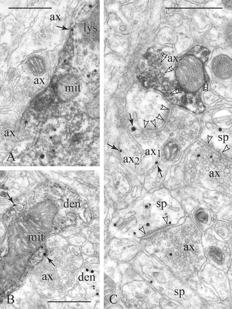
D1Rs in PV (diffuse precipitate) cellular profiles. A, PV perikarya display D1Rs in association with endomembranes, whereas only a fraction is expressed on the plasmalemma (arrow). B, Similarly, in PV dendrites (den), D1Rs may appear intracellularly and on nonsynaptic membranes (arrows). Note a second D1R-immunoreactive dendrite shown to the bottom right in B. C, A PV varicosity (double arrowheads point to synaptic vesicles) lies next to an e-l bouton [axon 1 (ax1)] forming a synapse with perforated postsynaptic density (between arrowheads). Unlike the PV axon, ax1 and neighboring ax2 show perisynaptic and extrasynaptic D1R labeling (arrows). Two other synaptic pairs also express D1Rs. mit, Mitochondrion; lys, lysosome; sp, spine. Scale bars, 400 nm.