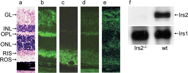
Figure 1.
Irs2 expression in the retina of adult WT mice. a, Eyes were collected from 9-week-old mice, and frozen sections were prepared for immunofluorescent staining (original magnification, 25×). a, WT retina with H&E staining; b, WT retina with antibody against Irs2; c, WT retina with anybody against rhodopsin; d, WT retina without primary antibody against Irs2; e, Irs2-/- retina with antibody against Irs2. f, The retinas were dissected from 9-week-old WT and Irs2-/- mice, and whole retinal lysates (50 μg) were separated by 7.5% SDS-PAGE gels. Western blotting was used to detect Irs1 and Irs2. GL, Ganglion cell layer; RIS, rod inner segments; ROS, rod outer segments.

