
Figure 5.
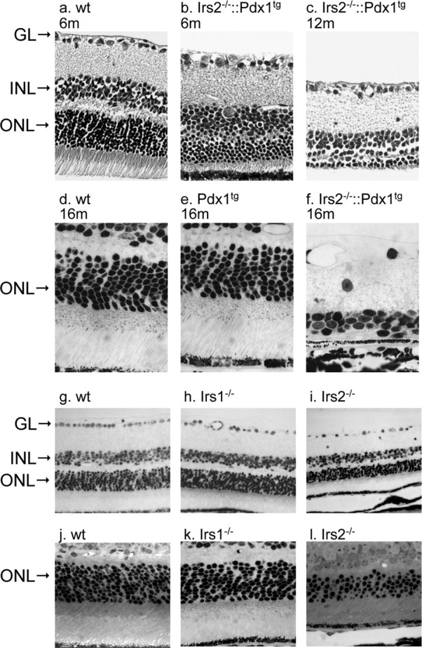
Morphological changes of retina in adult Irs2-/- and Irs1-/- mice. Eyes were collected from older Irs2-/-::Pdx1 at 6, 12, and 16 months of age, and cross sections were prepared for light microscopic observation (3 mice at 6 and 12 months, respectively; 7 mice at 16 months; a-f). g-l, Retinal cross sections obtained from 9-week-old WT, Irs1-/-, and Irs2-/- mice (original magnification is 20× for a, b, c, g, h, i and 40× for d, e, f, j, k, l). GL, Ganglion cell layer.