
Figure 6.
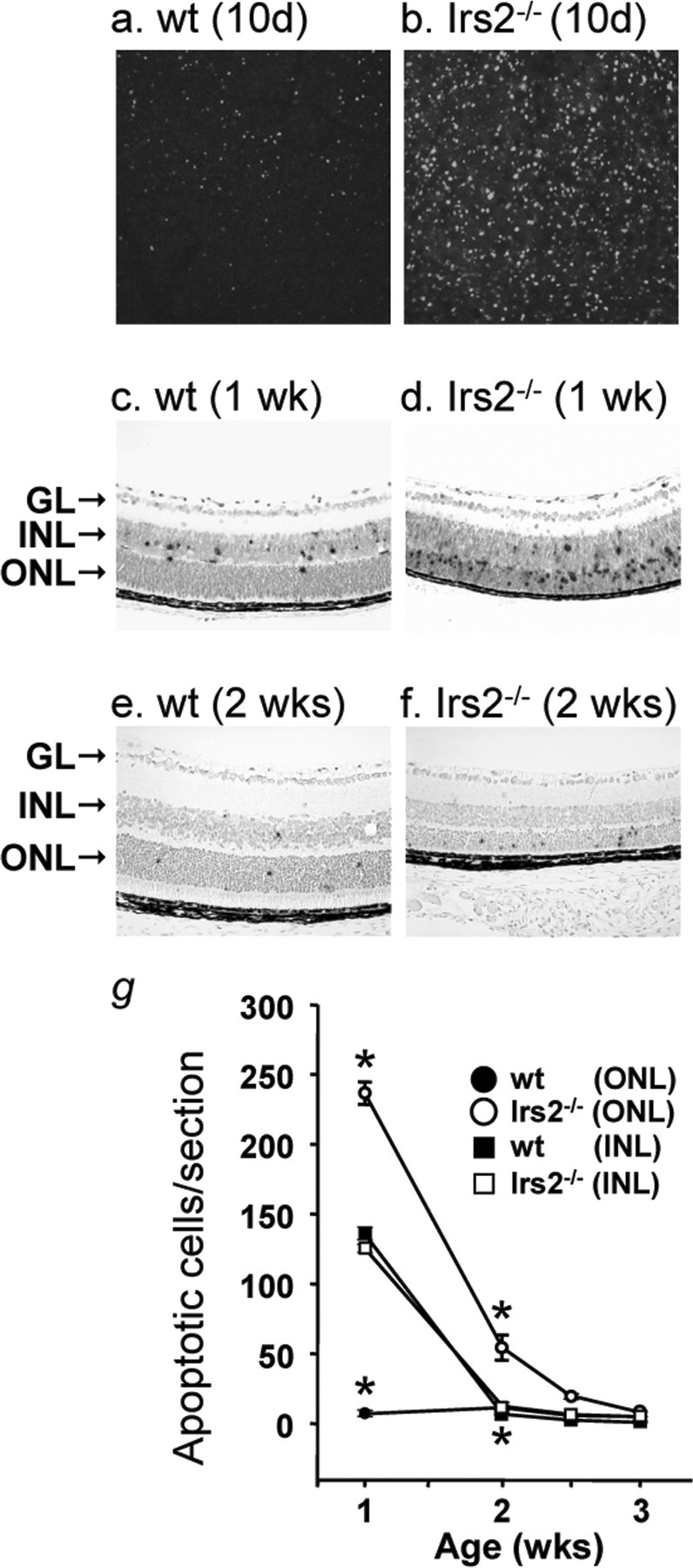
Apoptosis in the retina of Irs2-/- mice. TUNEL staining in whole-mount retina from 10-d-old WT and Irs2-/- mice using fluorescent method (a, b) (original magnification, 20×); TUNEL assay in paraffin retinal cross sections of 1- and 2-week-old WT and Irs2-/- mice using peroxidase method (c-f) (original magnification, 20×). Positive nuclei were counted and plotted as a function of age. Six retinal cross sections were measured and averaged from each mouse; data points indicate the average ± SD of three mice (g). GL, Ganglion cell layer. *p < 0.01.