
Figure 1.
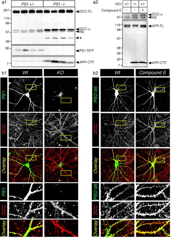
PS-dependent proteolysis of DCC in neurons. a1, Endogenous DCC-FL and proteolytic fragments (DCC-α and DCC-*) were detected by immunoblotting detergent lysates from Wt (PS1+/-) and KO (PS1-/-) mouse embryonic brains harvested at E15. A nonspecific band (NS) was also detected by this antibody. Bottom panels represent immunoblot analysis of PS1-NTF and APP-CTF. a2, In primary cultured neurons, although endogenous DCC expression was low, treatment with the γ-secretase inhibitor Compound E caused accumulation of DCC-α fragment to levels found in KO neurons. The bottom panel shows that endogenous APP-FL and APP-CTF were detected in untreated neurons (lane 1), KO neurons (lane 2), or neurons treated with Compound E (lane 3). Molecular weight standards are indicated on the left. b, Neuronal cultures were also immunostained with monoclonal antibody directed against DCC and polyclonal antibodies directed against PS1 (b1) or PSD-95 (b2). Representative confocal images are shown as a summation of contiguous three-stack images. The bottom panels show enlarged overlay images of the dendritic area. The level of overlap between two antibodies is shown as yellow.