
Figure 8.
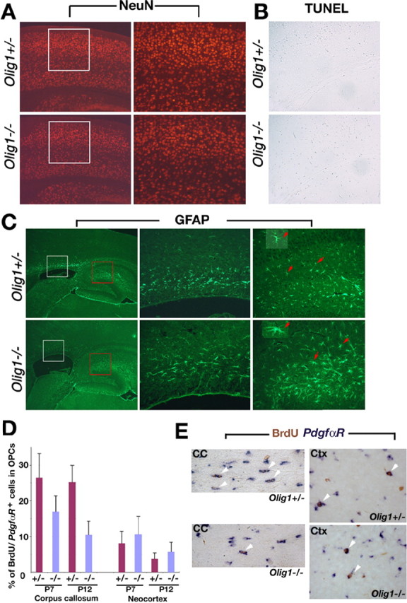
Neuronal development in brain from Olig1-null mice. A, NeuN expression in coronal sections of cortex from Olig1-heterozygous (+/-) and -null (-/-) mice at P14 to examine neuronal development. White-boxed areas in left panels are shown at higher power (20×) in the right panels. Neurogenesis and lamination are comparable in +/- and -/- mice. B, TUNEL of coronal sections from cortex indicates that there are few apoptotic cells in +/- and -/- mice. C, Astrocyte development examined in parasagittal brain sections using GFAP immunocytochemistry. White-boxed (corpus callosum) and red-boxed (hippocampus) areas (left panels) are shown at higher power (20×) in middle and right panels, respectively. GFAP+-reactive astrocytes (arrows) from -/- mice exhibit large cell bodies and bulky processes (bottom inset); however, astrocytes (arrows) from control mice have thin processes and relatively small cell bodies (top inset). D, The numbers of cycling cells in brain measured by double in situ hybridization and immunohistochemistry 4 h after BrdU administration to animals at P7 and P12. The proportion of PdgfαR+ OPCs in the cell cycle at the time of the BrdU injections in cortex is comparable between +/- and -/- mice but reduced in white matter from the null mutants. E, Examples of BrdU+/PdgfαR+ cells (arrowheads) are shown in the white matter and the cortex of Olig1 +/+ and -/- mice. CC, Corpus callosum; Ctx, cortex.