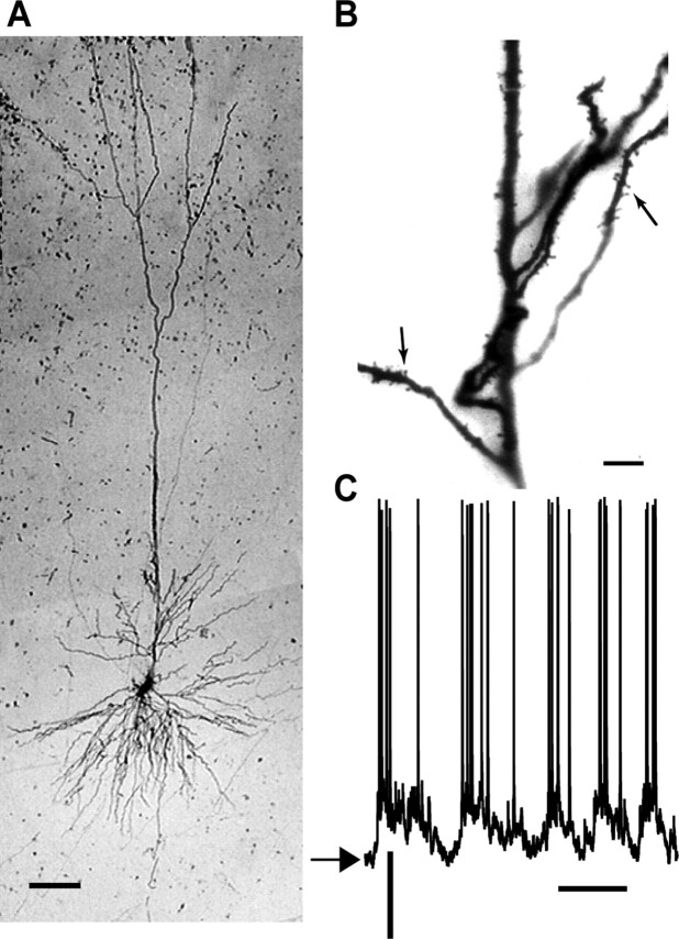
Figure 2.
Morphology and spontaneous activity of A1 neuron. A, A biocytin-filled pyramidal neuron. Scale bar, 100 μm. B, A section of its dendritic arborization, with dendritic spines clearly observable (arrows). Scale bar, 20 μm. C, Spontaneous membrane potential activity of the neuron. Resting potential is -75 mV (arrow). Calibration: 20 mV, 200 ms.

