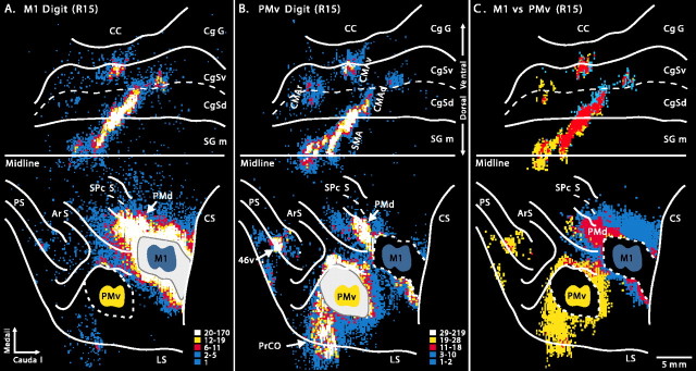
Figure 4.
Frontal lobe input to the digit representations of M1 and the PMv (R15). A, Location and density of neurons labeled after FB injections into the digit representation of M1. B, Location and density of neurons labeled after DY injections into the digit representation of the PMv. C, Overlap of the input to the digit representations of the PMv and M1. Red shading (overlap bins) indicates bins that have a high density of cells projecting to each injection site. Blue shading represents bins that have a high density of cells projecting to M1. Yellow shading represents bins that have a high density of cells projecting to the PMv. Abbreviations and conventions are as in Figures 1 and 2.

