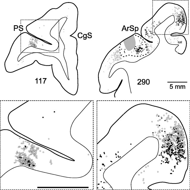
Figure 8.
Prefrontal and SMA neurons that project to the digit representations of the motor areas on the lateral surface. Left, Labeled neurons in area 46 that project to the PMv (open circles) or to M1 (filled circles) are plotted on a coronal section through the caudal third of the principal sulcus (R15). Right, Labeled neurons in the SMA that project to the PMv (open circles) or to the PMd (filled circles) are plotted on a coronal section located just caudal to the genu of the arcuate sulcus (R24). The FB injection site in the PMv is delineated by gray shading, and the surrounding region of intensely labeled neurons is delineated by the dotted line. CgS, Cingulate sulcus; PS, principal sulcus; ArSp, spur of the arcuate sulcus. Calibration, 5 mm.

