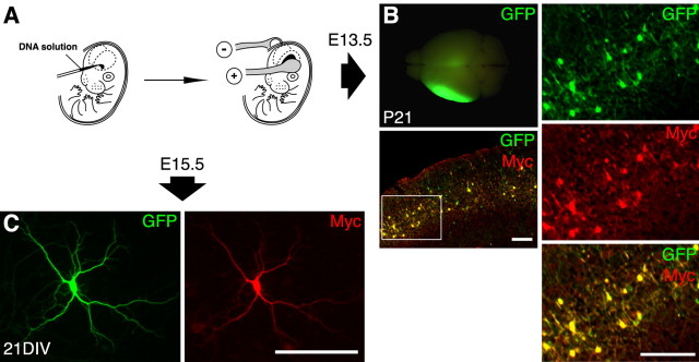
Figure 1.
The in utero electroporation procedure and its application in this study. A, Scheme of the in utero electroporation procedure. Expression vectors were injected into the left lateral ventricle of embryonic brains (left) and transfected into neuronal progenitor cells by in utero electroporation (right). B, GFP fluorescence in a rat brain (at P21) cotransfected with expression vectors encoding GFP and Myc-tagged PSD-Zip70C (left, top). A merged image of the thin section of the forebrain at P21 was double immunostained for GFP and Myc (left, bottom). High-magnification views of the segment enclosed in a white box are shown in the right column. C, Immunolabeling for GFP (left) and Myc (right) in cultured neurons (21 DIV) from the cerebral regions electroporated with GFP and Myc-tagged PSD-Zip70C expression vectors. Scale bars, 200 μm.

