
Figure 3.
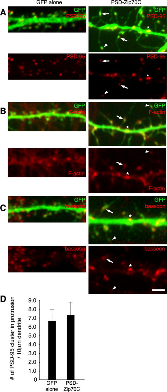
Characterization of PSD-Zip70C-induced elongated spines. Neurons transfected with GFP alone (left) or GFP and PSD-Zip70C (right) at 21 DIV were immunostained for GFP and PSD-95 (A), F-actin (B), or bassoon (C). The small head of thin spines (arrowhead), midbody of long filopodia (arrow), and shaft spine (asterisk) are indicated. D, Quantitative analysis of the number of PSD-95 clusters in protrusions per 10 μm dendrite from neurons transfected with GFP alone or GFP and PSD-Zip70C (each sample was quantified along a 100 μm dendrite of one neuron, and the number of PSD-95 clusters in protrusions was expressed as PSD cluster per 10 μm dendrite; n = 5 neurons) (mean ± SD). Scale bar, 2 μm.