Figure 6.

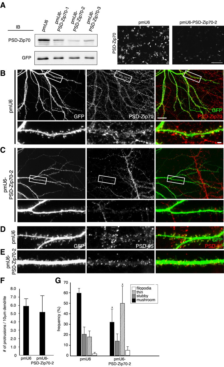
Effect of PSD-Zip70 knock-down on spine morphology. A, COS-7 cells cotransfected with GFP and Zip70WT expression vectors and pmU6 or pmU6-Zip70-1 to pmU6-Zip70-3 vectors were analyzed by immunoblotting (IB, left) and immunostaining (right) for GFP and/or PSD-Zip70. Scale bar, 200 μm. B-E, Hippocampal neurons (21 DIV) cotransfected with GFP and pmU6 or pmU6-Zip70-2 were double stained for GFP and PSD-Zip70 or PSD-95. F, G, Quantification of the spine density (number of spines per 10 μm dendrite length) (F) and morphology (G) (each sample was quantified with 1000 protrusions from 10 neurons; n = 10 experiments). Mean ± SD; *p < 0.05 compared with GFP alone (Student's t test). Scale bars: B, bottom, 2 μm; B, top, 20 μm.
