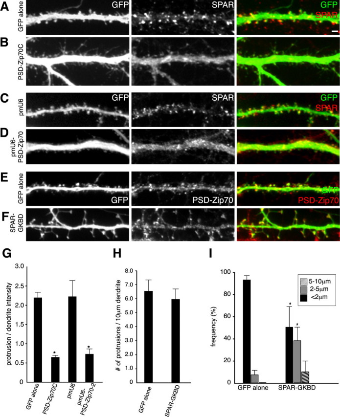
Figure 9.

Significance of PSD-Zip70 and SPAR interaction in spine morphology. The GFP expression vector alone (A) or GFP and PSD-Zip70C (B) were overexpressed in cortical neurons (21 DIV) by in utero electroporation. Neurons were immunostained for GFP or SPAR. The GFP expression vector and pmU6 (C) or pmU6-PSD-Zip70-2 (D) were cotransfected into hippocampal neurons (21 DIV) by microinjection. Neurons were immunostained for GFP or SPAR. GFP alone (E) or GFP and SPAR-GKBD (F) were overexpressed in hippocampal neurons (21 DIV) by microinjection. Neurons were immunostained for GFP or PSD-Zip70. G, The localization of SPAR in dendritic protrusions expressed as ratios of the fluorescence intensity of protrusion to that of shaft from neurons transfected with GFP alone, GFP and PSD-Zip70C, GFP and pmU6, or GFP and pmU6-PSD-Zip70-2 (each sample was quantified with 500 protrusions from 5 neurons). H, I, Quantitative analyses of the density (H) or length (I) of protrusions from neurons expressing GFP alone or GFP and SPAR-GKBD (each sample was quantified with 800 protrusions from 8 neurons; n = 10 experiments). Mean ± SD; *p < 0.05 compared with GFP alone (Student's t test). Scale bar (in A), 2 μm.
