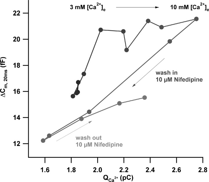
Figure 4.
Direct comparison of the effects of changes in single-channel current and open-channel number on RRP exocytosis. Scatter plot of ΔCm, 20 ms versus the corresponding Ca2+ current integrals of a representative perforated-patch experiment during (1) slow change of [Ca2+]e from 3 to 10 mm, (2) application of 10 μm nifedipine at 10 mm [Ca2+]e, and (3) wash out of nifedipine in the continued presence of 10 mm [Ca2+]e are shown. Data were smoothed by nonoverlapping two-point box car averaging. The time course of the experiment is indicated by the black-to-gray gradient.

