
Figure 1.
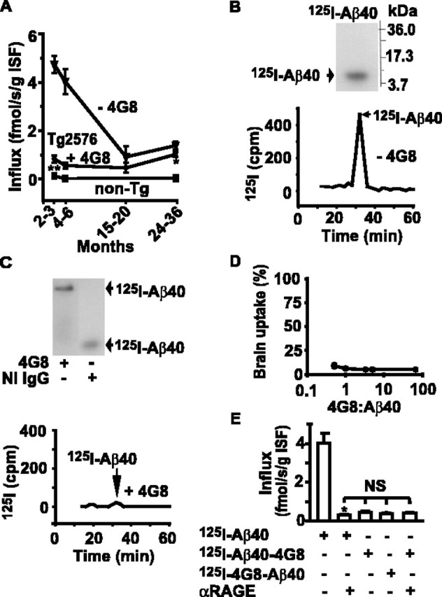
Aβ-specific IgG (4G8) blocks BBB influx of circulating Aβ40. A, Aβ40 BBB influx in APPsw+/– (Tg2576) mice in the presence (circles) and absence (triangles) of a fourfold excess of 4G8 determined with the brain perfusion method (LaRue et al., 2004) with 125I-Aβ40 at carrier concentrations corresponding to Aβ40 plasma levels in Tg2576 mice at different ages (Kawarabayashi et al., 2001), i.e., 4 nm (2–3 and 4–6 months), 3 nm (15–20 months), and 1.5 nm (24–36 months), and in age-matched littermate controls (non-Tg) at physiological Aβ40 plasma levels (50 pm) without 4G8. B, Tris-tricine SDS-PAGE (top) and HPLC (bottom) analysis of 125I-Aβ40 radioactivity in the arterial inflow without 4G8. C, Tris-tricine native PAGE (top) and HPLC (bottom) analysis of 125I-Aβ40 radioactivity in the arterial inflow with a four foldexcess of 4G8 or NI IgG. D, 125I-Aβ40 brain uptake at varying 4G8/Aβ40 plasma ratio in 4- to 6-month-old Tg2576 mice expressed as the percentage of control uptake. E, Transport across the BBB of 125I-Aβ40 and Aβ40–4G8 complexes labeled on either Aβ40 or 4G8 in the absence and presence of RAGE-specific IgG F(ab′)2 in 4- to 6-month-old Tg2576 mice. Mean ± SEM; n = 3–6. *p < 0.001; and **p < 0.05; NS, not significant.