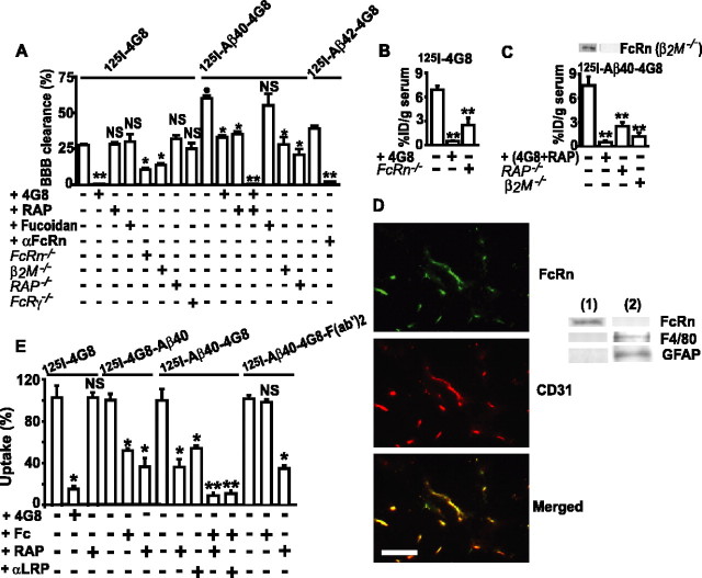
Figure 3.
FcRn-mediated BBB transcytosis of 125I-4G8, 125I-Aβ40–4G8, and 125I-Aβ42–4G8 from brain to blood. A, The BBB clearance of 40 nm 125I-4G8, 125I-Aβ40–4G8, or 125I-Aβ42–4G8 within 30 min of brain ISF microinjections with and without unlabeled 4G8 (2 μm), RAP (5 μm), fucoidan (1.5 mm), αFcRn (anti-FcRn, 60 μg/ml), and Aβ40 (1 μm) and in FcRn–/–, β2M–/–, RAP–/–, and FcRγ–/– mice. B, C, Serum levels of 125I-4G8 (B) and of 125I-Aβ40–4G8 (C) from experiments in A expressed as the percentage of injected dose (%ID). Western blot analysis of FcRn in brain microvessels in control and β2M–/– mice (inset, C). D, FcRn/CD31 double immunostaining in brains of nontransgenic mice (left) Scale bar, 50 μm. Western blot analysis (right) of FcRn, F4/80 (phagocytic cells), and GFAP (astrocytes) in isolated brain microvessels (lane 1) and capillary-depleted brain (lane 2). E, Uptake of 125I-4G8, 125I-4G8–Aβ40, 125I-Aβ40–4G8, or 125I-Aβ40–4G8-F(ab′)2 (1 nm) at the abluminal side of isolated mouse brain microvessels within 1 min at 37°C. 4G8 (5 μm), RAP (5 μm), Fc (10 μm), and αLRP (20 μg/ml) were applied as potential inhibitors. Mean ± SEM; n = 3–5. *p < 0.05, **p < 0.001, inhibitors or gene deletion versus the corresponding controls; •p < 0.01, 125I-Aβ40–4G8 versus 125I-4G8; NS, not significant.

